
BACK
Preserving Single-Cell Transcriptomic Signals of Mitochondrial Dysfunction in Cutaneous Lupus Erythematosus
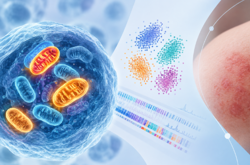
Mitochondrially encoded (MT) genes are treated nearly universally as quality-control nuisance and removed at a fixed threshold in single-cell RNA sequencing (scRNA-seq) analysis, yet the same transcripts carry critical information about cellular energetics and stress. Work in oncology has shown that discarding cells on the basis of high mitochondrial content can systematically remove viable, metabolically relevant populations rather than debris [1], and new probabilistic tools now make adaptive, cluster-aware filtering practical [2].
Systemic lupus erythematosus is a disease in which mitochondrial pathology is considerably implicated, yet this reassessment of mitochondrial filtering has not, to our knowledge, been extended to this research domain. A preliminary comparative analysis of single-cell transcriptomic signals with and without MT gene filtration in cutaneous lupus erythematosus (CLE; contrasting lesional vs. non-lesional skin) and healthy controls was performed. Retaining MT genes preserved a signal from Complex I mitochondrial subunits, a principal site of mitochondrial reactive oxygen species (ROS) production, in diseased skin and would have been lost with conventional filtering. This article sets out the rationale, the observation, and what its probable implications for scRNA-seq pipeline design in autoimmune disease.
Mitochondrial transcripts: artefact or signal?
A high fraction of mitochondrial reads has long been used as a proxy for cell damage. The reasoning is mechanical: when a cell’s plasma membrane is compromised during dissociation, cytoplasmic mRNA escapes while transcripts enclosed within intact mitochondria are retained, inflating the mitochondrial proportion of the remaining library. Most pipelines therefore apply a fixed cut-off, commonly 5%, 10%, or 15% of counts, and discard everything above it.
The limitation of this conventional approach is that mitochondrial content is not constant across cell types or tissues. Cardiomyocytes, renal tubular cells, activated lymphocytes, and metabolically reprogrammed tumour cells all carry a legitimately high mitochondrial fraction. A single global threshold cannot separate a dying cell from a respiring one. Yates and colleagues demonstrated exactly this in cancer scRNA-seq, showing that standard mitochondrial filtering depletes viable malignant populations with altered metabolism, biasing the surviving dataset [1].
Several developments, two of which are most relevant to ThinkBio’s efforts, have made it possible to act on this limitation. The first is adaptive thresholding. MitoChontrol, a recently described probabilistic framework, models the mitochondrial transcript fraction within transcriptionally coherent clusters as a Gaussian mixture and sets a per-cluster threshold at the point where the posterior probability of cellular compromise exceeds a user-defined confidence level. Applied to a pancreatic ductal adenocarcinoma dataset, it removed compromised cells while retaining biologically elevated but viable populations, outperforming fixed-threshold and outlier-based approaches [2].
The second is annotation. Because mitochondrially encoded genes are routinely stripped during quality control, conventional enrichment analyses against general-purpose resources rarely recover mitochondrial biology in any interpretable form. MitoCarta3.0 addresses this directly, providing a curated inventory of 1,136 human mitochondrial genes with sub-organelle localisation and assignment to 149 hierarchical MitoPathways [3]. Adaptive filtering and curated mitochondrial pathway annotation are complementary: the first preserves the signal, the second makes it biologically interpretable.
Lupus as the test case
This literature is predominantly built in cancer. Autoimmune disease is where we would expect it to generalise most readily, and lupus would be an ideal candidate.
Mitochondrial pathology in lupus is not incidental. Altered bioenergetics and oxidative stress across immune cell subsets are established contributors to disease mechanism and have been proposed as clinical readouts [4]. Heightened mitochondrial metabolism and oxidative stress promote the escape of mitochondrial nucleic acids into the cytosol, where they are sensed as damage-associated molecular patterns and drive type I interferon and inflammasome activation across lupus manifestations [5], including cutaneous lupus erythematosus (CLE) [6].
Analytical approach
Single-cell transcriptomic signals derived from the same CLE dataset were processed in two ways: under conventional fixed-threshold mitochondrial filtering, and with mitochondrially encoded genes retained. Mitochondrial genes were then examined in the context of processes characteristic of lupus, particularly oxidative stress [4].
This was a preliminary, hypothesis-generating comparison rather than a detailed benchmarking study. Its purpose was to establish whether a mitochondrial signal may survive in CLE that standard quality control would otherwise remove.
Observation
Retaining mitochondrial genes preserved a coherent signal from mitochondrial Complex I subunits (the MT-ND family), which were upregulated in diseased skin relative to healthy tissue. Complex I is a principal generator of mitochondrial reactive oxygen species, and its transcriptional elevation in diseased tissue is consistent with the mitochondrial stress described in lupus pathogenesis [4]. Under conventional fixed-threshold filtering, this signal was lost.
The finding is preliminary and requires validation in larger, independent cohorts. It is nonetheless a concrete demonstration that the mitochondrial fraction in CLE scRNA-seq is not purely technical noise, and that the routine decision to discard it comes with a cost.
Implications for pipeline design
We do not propose that mitochondrial genes should simply be retained. Doing so would reintroduce exactly the artefact the threshold was designed to control, and dying cells remain a real contaminant. The useful conclusion is finer and more actionable: mitochondrial filtering should be treated as a tunable analytical decision with biological consequences, not as a fixed preprocessing default. Two strategies follow.
Adjunct mitochondrial analysis. Mitochondrial transcripts can be analysed as a dedicated layer alongside the conventionally filtered dataset, preserving standard quality control for the primary analysis while extracting mitochondrial biology separately. This mirrors the approach taken in cancer genomics, where mitochondrial content has been treated as an informative axis in its own right- as in the demonstration that respiratory complex and tissue lineage shape recurrent mutations in tumour mtDNA [5].
Adaptive, cluster-aware thresholding. Alternatively, per-cluster probabilistic thresholds of the kind implemented in MitoChontrol [2] can be adopted in place of a global cut-off, retaining metabolically active cells while still excluding compromised ones. Paired with MitoCarta-based pathway annotation [3], this supports functional inference from transcripts that would otherwise never reach the analysis.
Conclusion
The same analysis applied to larger cohorts, and across other manifestations of lupus, systemic, renal and cutaneous, would establish whether the Complex I signal observed here is a general feature of lupus tissue or specific to skin. Extending it to other immune-mediated inflammatory diseases, where mitochondrial dysfunction is similarly implicated, would be the obvious next step.
There is also a downstream consideration that is difficult to ignore. Single-cell transcriptomic foundation models are trained on data that has already passed through conventional quality control, which means that whatever fixed mitochondrial thresholds remove is absent from the representations these models learn. If mitochondrial signal is systematically discarded upstream, no amount of model capacity recovers it. Improving how mitochondrial content is handled at the quality-control stage is therefore not only a matter of analytical hygiene; it directly affects the signal-to-noise ratio available to the next generation of single-cell models.
Implementing this does not require rebuilding an existing pipeline. A workable pattern for lupus datasets is to keep standard quality control for the primary analysis object, but to retain the mitochondrial count matrix rather than discard it at ingestion; to replace the global percentage cut-off with a per-cluster probabilistic threshold so that metabolically active immune populations are not removed before clustering has even happened; and to carry mitochondrially encoded genes forward as an annotated layer, scored per cell and per cluster against MitoCarta MitoPathways [3] alongside interferon and other disease-relevant signatures. The practical shift is one of framing: the mitochondrial fraction becomes a covariate to be modelled and interpreted rather than a filter to be applied once and forgotten.
The argument for doing this across autoimmune disease is mechanistic, not merely methodological. Wherever mitochondrial nucleic acid release, oxidative stress, and altered bioenergetics are part of how a disease operates, the mitochondrial content of a cell’s library is in part a readout of that pathology rather than a measure of how well the sample was handled.
At ThinkBio, mitochondrial handling is one component of a broader effort to make single-cell and bulk transcriptomic pipelines interpretable at the level of patient biology. Work of this kind, questioning defaults that have hardened into convention, is where much of the recoverable signal in existing datasets still sits untapped.
References
1. Yates J, Kraft A, Boeva V. Filtering cells with high mitochondrial content depletes viable metabolically altered malignant cell populations in cancer single-cell studies. Genome Biol. 2025;26:91. doi:10.1186/s13059-025-03559-w
2. Strassburg C, Pitlor D, Singhi AD, Gottschalk RA, Uttam S. MitoChontrol: adaptive mitochondrial filtering for robust single-cell RNA sequencing quality control. bioRxiv [Preprint]. 2026. doi:10.64898/2026.04.04.716517
3. Rath S, Sharma R, Gupta R, et al. MitoCarta3.0: an updated mitochondrial proteome now with sub-organelle localization and pathway annotations. Nucleic Acids Res. 2021;49(D1):D1541–D1547. doi:10.1093/nar/gkaa1011
4. Yennemadi AS, Keane J, Leisching G. Mitochondrial bioenergetic changes in systemic lupus erythematosus immune cell subsets: contributions to pathogenesis and clinical applications. Lupus. 2023;32(5):603–611. doi:10.1177/09612033231164635
5. Gorelick AN, Kim M, Chatila WK, La K, Hakimi AA, Berger MF, Taylor BS, Gammage PA, Reznik E. Respiratory complex and tissue lineage drive recurrent mutations in tumour mtDNA. Nat Metab. 2021;3(4):558–570. doi:10.1038/s42255-021-00378-8
6. Klein et al.Epidermal ZBP1 stabilizes mitochondrial Z-DNA to drive UV-induced IFN signaling in autoimmune photosensitivity.Sci. Immunol.10,eado1710(2025).DOI:10.1126/sciimmunol.ado1710


Build the future of
biology with us
Contact Us
Stay Ahead with
ThinkBio.Ai®
Subscribe to our newsletter to receive the latest updates on products, sustainability efforts and services.