Spatial Omics – The Need for High-Resolution Diagnostics in Immunotherapy
Cancer immunotherapy has revolutionized oncology, offering the possibility of long-term control or even complete eradication of disease in some patients. Among these therapies, immune checkpoint inhibitors (ICIs) have achieved the greatest success in the treatment of solid tumors, dramatically improving outcomes across various cancer types.
However, the reality remains that a substantial proportion of patients either do not respond to ICIs or experience serious adverse effects. This unpredictability highlights a critical unmet need, a better predictive tool and a deeper mechanistic understanding of how these therapies work and why they sometimes fail.
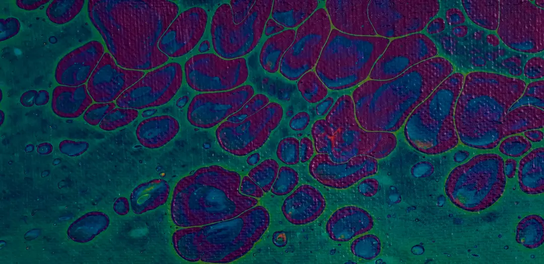
This is where spatial omics technologies are emerging as game changers. Spatial omics is the analysis of high-resolution whole tissue images. By providing insights into the spatial organization of cells and molecules within the tumor microenvironment, spatial omics promise to enhance our understanding of cancer biology and lead to more personalized, effective treatments.
Introduction to Spatial Omics
Spatial omics is a cutting-edge technology that maps positional relationships between cells within a tissue. Consider for example, a tumor tissue, composed of five major cell types – tumor epithelial cells, many different immune cells, stromal cells, endothelial cells and normal epithelial cells. Spatial mapping identifies the location of each and every cell-type, and how they are organized with respect to each other. This can help understand cancer progression and one can generate genomics and proteomics data on top of the spatial data to create a readout of each cell. The readouts can be gene expression (spatial transcriptomics), protein expression (spatial proteomics) and so on.
This high-resolution mapping of tumors reveals the intrinsic heterogeneity of the tissue, which is often highly variable from one area to another. Variability comes not only from the cellular composition but also how cells are organized with respect to each other.

Key differences between spatial omics and other omics platforms
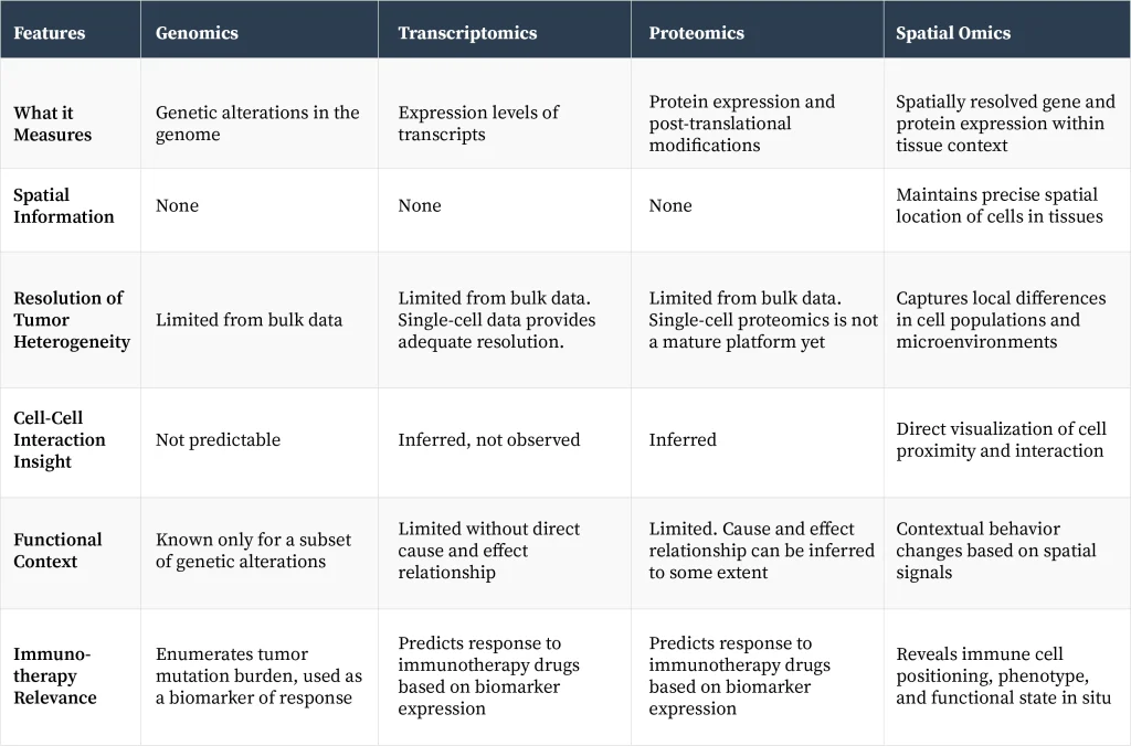
Understanding these differences can reveal why some therapies work in certain patients but not in others, why some patients’ tumors spread, while for others the tumor is localized? These insights help clinicians to manage the disease more efficiently, and drug developers use this knowledge to develop novel therapies.
Spatial Omics has the potential to make Cancer Immunotherapy more effective
Cancer immunotherapy relies on body’s own immune cells to attack and eliminate tumors. Therefore, prior knowledge of how immune cells are organized in different regions of tumor tissue and the composition of immune cells in different regions of the tumor is crucial for cancer immunotherapy to work. Spatial omics has addressed some of the most pressing challenges in cancer immunotherapy:
- Predictive Insights: Detailed spatial maps can quantize the presence of different immune cells in the tumor microenvironment. It also uncovers functional patterns in these cells, which can inform whether the tumor will respond to treatment or remain unresponsive. Such knowledge can help to select patients who will benefit from the therapy. Given that cancer immunotherapy induces toxicity, unresponsive patients can be spared from adverse effects of the drug.
- Identification of New Therapeutic Targets: By revealing immune cell dynamics and tumor-immune interactions, spatial omics technologies can identify novel points of intervention. For example, in tumors that are immune deserts – meaning lack of infiltration of immune cells, a therapy to improve immune cell infiltration will make cancer immunotherapy drugs work more effectively.
- Tracking Tumor Evolution: Spatial analysis enables the study of tumor subclones and their distinct behaviors within different regions of the tumor mass. Sub clonal populations of cells harbor unique genetic alterations responsible for drug resistance and tumor relapse.
- Characterization of Immune Cell Behavior: Clonal expansion of B and T cells in different regions of the tumor and the identity of their receptors – B and T cell receptors, reveal the presence of unique immunogenic tumor antigens, which can contribute to the development of next-generation cancer vaccines and cell therapies.
- Beyond Conventional Biomarkers: Spatial technologies can reveal critical biological changes in the tumor microenvironment, including metabolic shifts and structural DNA alterations, that traditional diagnostics are likely to miss.
Collectively, these insights open to new refined therapeutic strategies, reduce unnecessary side effects, and ultimately improve patient outcomes and their quality of life.
ThinkBio Pixelomics™ Solution: The Pixelomics solution from ThinkBio provides an end-to-end AI pipeline to analyze spatial omics data. For the Spatial Proteomics solutions as part of Pixelomics, we use established models to segment the images, and specialized algorithms to do cell alignment, denoising and clustering of the cell images. The final step is classification of the cells as per the relevant biomarkers.
Challenges in Clinical Implementation
Despite its transformative potential, integrating spatial omics technologies into routine clinical practice presents significant obstacles:
- Cost and Turnaround Time: Spatial analyses remain expensive and time-intensive, limiting their feasibility for large-scale clinical deployment at present. High resolution data collection is especially expensive.
- Operational Complexity: Effective use of spatial technologies demands close collaboration among multidisciplinary teams, including clinicians, laboratory scientists, and data analysts — a logistical challenge for many institutions.
- Clinical Usability: Hospitals and clinics require diagnostic tests that are rapid, standardized, and easily interpretable, whereas spatial analyses currently involve complex procedures and specialized expertise.
- Understanding Systemic Effects: Immunotherapy exerts effects throughout the body, yet spatial technologies predominantly focus on the tumor site. Integration with systemic data, such as from blood or lymph nodes is necessary for a comprehensive interpretation.
- Causality vs. Correlation: While spatial mapping can identify associations, understanding the underlying biological mechanisms requires perturbational studies, including the development of new model systems like patient-derived organoids, which may not fully recapitulate the intrinsic features of a tumor.
What can we expect in the foreseeable future?
The applications of next-generation technologies to solve problems in human health, such as genome sequencing, identification of biomarkers by high throughput mass spectrometry and analysis of tumor microenvironment demonstrate that complex innovations can, over time, become integral to routine care if they are clinically impactful.
For spatial omics technology, the immediate opportunity lies in targeted application: Deploying high-resolution spatial analyses in selected patient groups where detailed insights can have direct clinical benefit, for example, a treatment strategy when first, second, and third-line therapies have failed. Faster clinical adoption and widespread use will come when the technology gets further refined to enhance speed, reduce cost, and simplify workflows.
Case Study
Let us look at a case study for one of the pipelines in Pixelomics – Spatial Proteomics.
Problem Statement: Analyze whole tissue images with cells, cell membranes and nuclei to classify cells as per specific biomarkers. The images are generated on multiple channels with each channel corresponding to a specific biomarker protein.
Dataset: We used the tonsil dataset, collected as part of the Cooperative Human Tissue Network project. The imaging technique used was Multiplex Immunohistochemistry (mIHC). There are 17 channels in this image.

Our AI Pipeline:

The final goal of this pipeline is to classify each cell as per their type and to cluster cells as per the cell types to allow the oncologist to see clustering of the tumor cells.
Segmentation: The first step in the pipeline is to segment the image into individual cells

Noise removal for CD66b channel as example: There is ambient noise in the images that needs to be improved.


Clustering with spatial overlay of each cluster: The final step in the pipeline is to cluster these cells based on their spatial position as well as their type. This enables the researcher to investigate the proliferation of the tumor.
