Introduction
Spatial omics is a next-generation approach that integrates molecular profiling with spatial context, enabling precise mapping of cell types and their organization within tissues. In tumors, where epithelial, immune, stromal, endothelial, and normal cells coexist, this technology provides a high-resolution view of the tumor microenvironment. By combining gene expression (spatial transcriptomics) and protein expression (spatial proteomics), spatial omics reveals the intrinsic heterogeneity of tumors and shows how cellular composition and spatial arrangement shape disease progression and therapeutic response.
ThinkBio®’s Pixelomics™ solution builds on this foundation with an end-to-end AI pipeline for spatial omics analysis. In spatial proteomics applications, Pixelomics™ employs advanced models for image segmentation, along with specialized algorithms for cell alignment, denoising, and clustering. The pipeline concludes with biomarker-based cell classification, transforming complex spatial datasets into actionable insights for research and clinical decision-making.
Let us look at a case study for one of the pipelines in Pixelomics™ – Spatial omics
Problem Statement: Analyze whole tissue images with cells, cell membranes and nuclei to classify cells as per specific biomarkers. The images are generated on multiple channels with each channel corresponding to a specific biomarker protein.
Dataset: We used the tonsil dataset, collected as part of the Cooperative Human Tissue Network project. The imaging technique used was Multiplex Immunohistochemistry (mIHC). There are 17 channels in this image.

Our AI Pipeline:

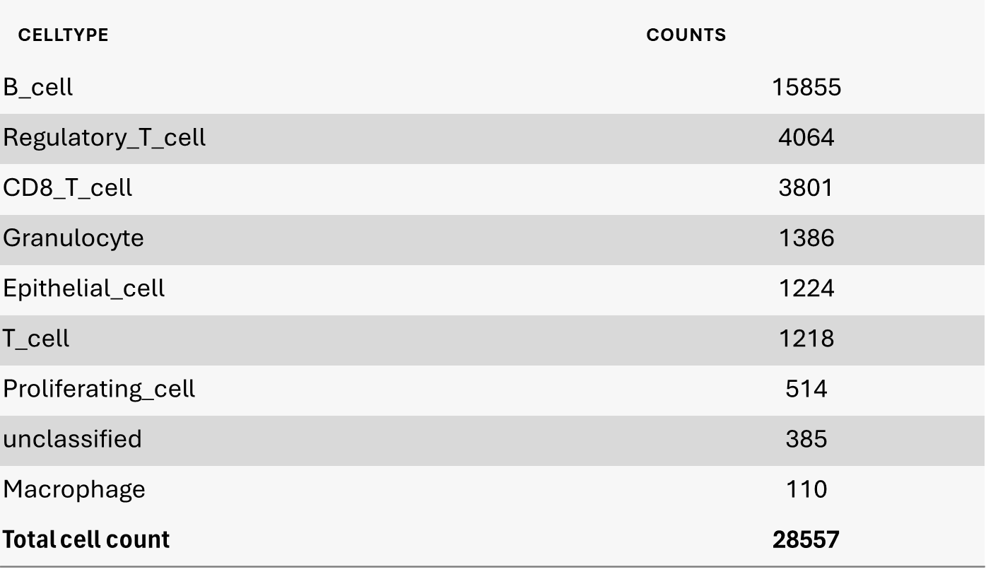
The final goal of this pipeline is to classify each cell as per their type and to cluster cells as per the cell types to allow the oncologist to see clustering of the tumor cells.
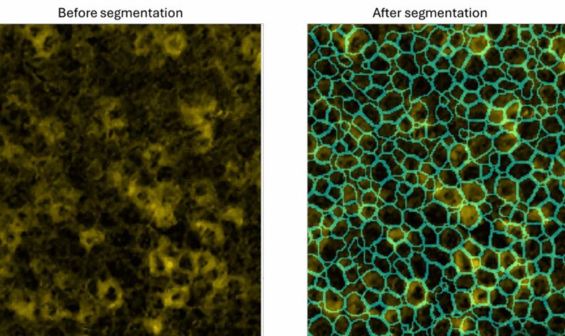
Segmentation:
The first step in the pipeline is to segment the image into individual cells
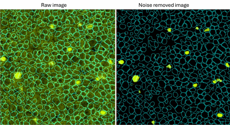
Noise removal for CD66b channel as example:
There is ambient noise in the images that needs to be improved.
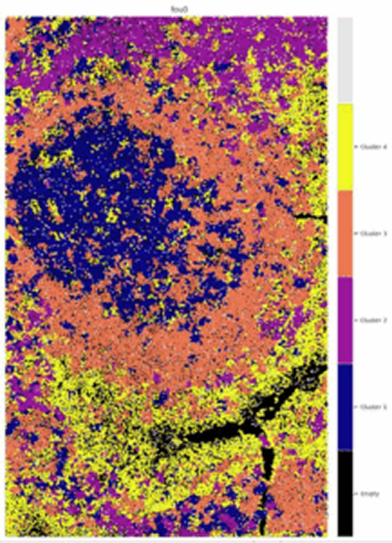
Clustering with spatial overlay of each cluster:
The final step in the pipeline is to cluster these cells based on their spatial position as well as their type. This enables the researcher to investigate the proliferation of the tumor.